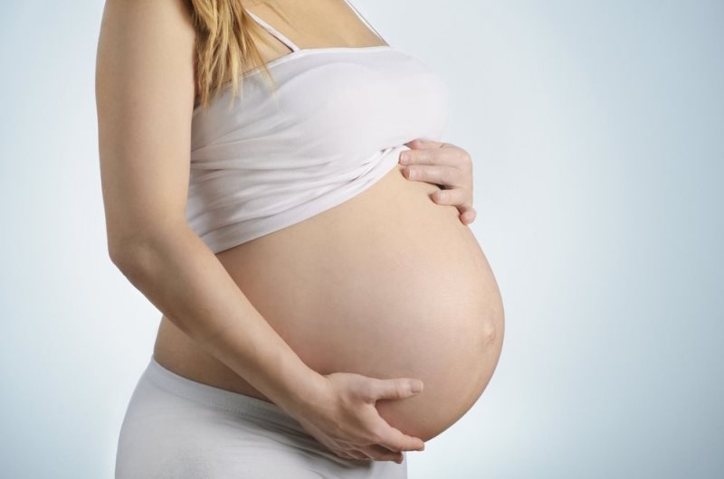
Cutaneous manifestations in pregnancy: Main dermatoses
A number of dermatological conditions are either identified as pregnancy-specific or more commonly occur during pregnancy. They may be confused due to their difference in terminology throughout different countries. Shornick (1998) concluded that only three diseases are universally accepted as being pregnancy-specific: gestational pruritus caused by cholestasis; pruritic urticaria papules and pregnancy plaques, as well as gestational herpes. The pathogenesis of pregnancy dermatoses, except for Herpes gestationis is still obscure and raises many questions that have not yet been answered. There are a series of generating factors, and attempts were made to split them into three categories: predisposing factors, contributing factors and determinants. These endogenous or exogenous risk factors may occur in isolation or in association with and are able to instigate the occurrence, recurrence or aggravation of these diseases. These include hormones like estrogen, progesterone; genetic predisposition; autoimmunity; sun exposure; age under 20 years; liver disease, malnutrition, anemia, hormonal disorders, infections, psycho-emotional condition of pregnant women.
The following classification system with different international differences in the nomenclature is presented:
- Pruritus gravidarum
- Polymorphic eruption of pregnancy (or papules and pruritic urticarial plaques)
- Prurigo of pregnancy (or gestational prurigo, papular dermatitis)
- Pruritic pregnancy folliculitis (or impetigo herpetiform)
- Gestational herpes (synonym – pemphigoid gestationis, bullous pemphigoid in pregnancy).
This classification does not include atopic eczema, which, according to some studies, includes half of the symptomatic skin diseases in pregnancy. Fortunately, inflammatory skin disorders such as atopic dermatitis or psoriasis have no significant effects on pregnancy evolution.
According to the another classification, there are four specific dermatoses of pregnancy:
- Pemphigoid gestationis
- Pruritic urticarial papules and plaques of pregnancy (PUPPP)
- Atopic eruption of pregnancy
- Intrahepatic cholestasis of pregnancy
Each pregnancy dermatosis is distinct and carries different risks for the mother and the fetus.
A distinction must be made between dermatological diseases that happen to occur while the patient is pregnant versus specific dermatoses that occur only during pregnancy. Pregnant patients with dermatologic conditions requiring treatment should be co-managed by the obstetrician and the dermatologist.
Pruritus
Complaints of pruritus during pregnancy are common and its incidence varies directly with the need for a medical healthcare.
Pruritus Gravidarum (Intrahepatic Cholestasis of Pregnancy)
This disease is considered a monosymptomatic form of intrahepatic cholestasis of controversial etiology. It is a rare disease with genetic predisposition. Is a reversible form of hormonal triggered cholestasis, which occurs in third trimester of pregnancy. Intrahepatic cholestasis tends to recur in subsequent pregnancies (45-70%). The incidence of intrahepatic cholestasis of pregnancy, in contrast to other dermatoses in pregnant women, presents a pattern dependent on the geographical area.
Incidence: it commonly occurs in 1-2% of cases.
Etiology: This skin disorder commonly occurs in pregnancy and is not a primary dermatological disease. It is considered a slight variation of intrahepatic cholestasis in pregnancy. Contributing factors are the hormones of pregnancy, genetic predisposition (18, 31 HLA antigens), dyslipidemia, and environmental factors.
Pathophysiology: In cholestasis, the serum level of bile salts is increased, which is then stored in the dermis, causing itching. Scratches and excoriations are present. Non-specific histological features. Primary skin lesions are missing.
Clinical features: It is characterized by an overwhelming feeling of scratching causing major skin lesions. The disease onset starts in the second half of the pregnancy evolution, commonly in the extremities and the abdominal region and generalizes relatively quickly. On a continuous basis, pruritus show periodic exacerbations, especially during the night under the influence of heat, in stressful or particular emotionally charged situations. It is usually accompanied by insomnia, irritability, physical and mental discomfort, fatigue, and leads to intense scratching, which in turn leads to secondary skin lesions, where itching is replaced by pain. Sometimes it is accompanied by jaundice. It is commonly characterized by absence of any primary skin lesion.
Diagnosis: As a specific marker of hepatocellular integrity, the serum concentration of glutathione S-transferase alpha was dosed and administered to patients who develop this disease. Disturbed excretion of biliary salts with increasing maternal serum values is considered to be the major factor of this disorder. However, the most steady diagnostic markers are characterized by an increase in total bile acid levels above 11 μmol / l, with a 42% increase in cholic acid and a decrease in glycine / taurine ratio below 1. Although bilirubin, transaminases, and alkaline phosphatase may be elevated, the hallmark of ICP is elevation of serum bile acids
Treatment: In local symptomatic treatment, talcum powder and menthol mixtures are used. Pathogenic treatment attempts to remove the consequences of intrahepatic cholestasis. Initially, cholestyramine (anion exchange resin – bile-binding acid) was administered 4g /4 times / day in tea, juice or milk. A series of studies have been conducted to prove that treatment with ursodeoxycolic acid 8-10mg / kg / day is superior to the administration of 8g / day of cholestyramine during 14 days.
Most publications recommend early induction of labor, commonly at 37 to 38 weeks. When cholestasis is severe, delivery is considered earlier if fetal lung maturity is established.Obstetric approach includes artificial interruption of pregnancy, avoiding premature birth and sometimes intrapartum follow-up.
Maternal prognosis is good, pruritus disappears spontaneously over 3-4 days postpartum, rarely it may lead to a process of liver cirrhosis or other severe chronic diseases. There is a risk of postpartum hemorrhage. The prognosis regarding the pregnancy evolution is associated with high occurrence of premature births (in over 44%). Poor fetal prognosis is mostly due to premature births, as well as other cases of intrauterine impairment (22%), intracranial hemorrhages and even exitus (1-2%).
Papules and pruritic urticarial plaques
Incidence: in 0.25-1% of cases. It is the commonest pregnancy dermatosis.
Etiopathogenesis: Not clear. There is no immunoglobulin or complement stored in the dermis, whereas the absence of the C3 linear band in the basement membrane helps to differentiate them from gestational herpes. It has been hypothesized that this disorder can be stimulated by fetal cells that invade the maternal skin. Furthermore, similar triggering mechanisms are proposed for lupus, systemic sclerosis, and postpartum thyroiditis.
Histopathology: Perivascular lymphocyte infiltration; the histopathological aspect is nonspecific with presence of eosinophilic component.
Clinical features: Eruptive lesions occur in the third trimester of gestation, commonly at first pregnancy. These can also occur in multigravidae, and sometimes even postpartum. It is more likely to be found in women pregnant with male fetus. They are rash-like, erythematous plaques, on which small papules (1-2 mm in diameter), surrounded by a small pale halo, develop. Papules whiten on vitropression. They develop first on the skin of the abdominal region, and then extend on the thighs, buttocks and arms.The eruption is rarely seen on the trunk, while the face is constantly affected. They are extremely pruritic and accompanied by scrathing-induced lesions.
Diagnosis: Routine investigations showed normal results. Negative direct and indirect immunofluorescence reactions.
Topical treatment is aimed at relieving pruritus by using soothing solutions, emollient products, moisturizing creams, antipruritic preparations of 0.1-1%, menthol, 0.5-2% camphor, 0.5-1.5% ichtiol, 0.5-2% phenol. Topical steroids represent the most valuable therapeutical approach, specifically the high-potency ones (0,1% triamcinolone acetonide or fluocinolone acetonide). Synthetized antihistamines (promethazine) are administered systemically, whereas corticotherapy (Prednisone) plays an important role in the treatment of this disorder, especially in severe cases.
Commonly it does not influence the maternal and fetal prognosis, as well as pregnancy evolution.
Prurigo pregnancy (Prurigo gestationis Besnier)
The disorder was initially described by Besnier, and was later included in the category of papulo-pruritic pregnancy dermatoses.
Incidence: rarely, (1: 300-1: 2400)
Etiopathogenesis: A strong component of atopic eczema.
Histopathology is nonspecific, perivascular lymphocytic infiltration is highlighted; parakeratosis, acanthosis; negative immunofluorescence reaction.
Clinical features: The disease onset occurs in the second half of the pregnancy, the skin eruption being preceded and accompanied by intense pruritus. The primary lesion is represented by a dermal papule of edematous type, often infiltrated and being centered by a small vesicle surrounded by an erythematous halo. However, it is rarely clinically detected since it is quickly damaged by scratching and replaced with a haematic crust. Lesions can be isolated or clustered and are predominantly and preferentially located on the flexor parts of the upper and lower limbs, on the dorsal part of thorax, and buttocks. They are accompanied by scrathing-induced lesions.
Diagnosis: The biological features are within normal limits and appropriate to the gestational condition and age at which it occurs. It has been observed to occur more frequently in women with atopic conditions and is associated with an increased level of IgE.
Treatment: The main purpose of local medication is to accelerate the healing of skin lesions and to eliminate or soothe the extremely annoying subjective symptoms (pruritus). Antipruritic lotions and mixtures containing: 1% menthol, 1-3% anesthetics, 1-2% phenol, 0,5-1,5% ichthyol, 1-5% chlorohydrate, 0,5-1% resorcinol, 0, 5% camphor, 2.5-5% lidocaine hydrochloride, antihistamines (Doxepin), and diphenhydramine.
The following antiseptics are used: hydrogen peroxide, 0.1% potassium permanganate, zinc sulphate as in Dalibor’s solution and / or Castellani tincture in cases of pyodermies. The best results were obtained when applying dermatocorticoids (lower potency) or in various combinations of them.
Maternal, pregnancy and fetal prognoses are good, since dermatosis is referred to the category of benign diseases.
Pruritic pregnancy folliculitis (or impetigo herpetiform)
The disease is considered an exudative form of psoriasis, with a particular evolution during pregnancy.
Incidence: rarely.
Etiopathogenesis: Unknown aetiology disorder.
Histology: characterized by sterile follicular papules, developing into acute folliculitis with mixed inflammatory cells, upper spherical edema, but negative for germs. Microabcesses; Kogoj’s spongiform pustules; neutrophils.
Clinical features: It occurs in the second or third trimester of pregnancy. It is considered an exudative form of psoriasis, with a particular evolution during pregnancy. It occurs in young primiparous women in the second trimester of gestation. It shows a low incidence. The disease onset is presented with fever, chills, dry tongue, sweating, vomiting, diarrhea, frequent delirium, convulsions, tetany, nystagmus, dysphagia. The skin eruption occurs along with general phenomena or after a few hours. It is commonly located on the abdomen, the genito-crural region, submammary ditches, the axillary regions, rarely on face, neck or knees. Subsequently, it covers the entire skin surface except for palms. This eruption is presented by round, oval or polycyclic erythematopoietic plaques, on which numerous superficial pustules are formed and clustered into herpetiform bunches. As the disease develops, the pustules extend to the peripheral side of the plaque, whilst the center remains erythematosus-crustaceous, followed by a continuous process of pustulization. No ulcers or scars occur. Sometimes the Kobner phenomenon (a skin reaction involving isomorphic reproduction of the lesions characteristic for a traumatised area) can be observed. The skin is eroded at folding regions, exudative, sometimes vegetative. The mucous membranes are often involved, especially the buccal one where microulcerations occur, but these can also affect the digestive tract. Appendages are also affected by the appearance of alopecia and nail lesions, but these are secondary to rash. Subjectively, the feeling of pruritus is associated with pain, and if full-blown, it becomes annoying, since every movement, whether voluntary or not, is accompanied by a sensation that the skin cracks. The overall condition changes as well. The silent evolution of the disease can be confused with erythrodermic psoriasis characterized by red skin and covered by a very thin epidermis that continuously exfoliates.
Diagnosis: negative direct immunofluorescence reaction.
Treatment: Currently, the systemic treatment of dermatosis is based on the use of corticosteroid therapy: Prednisone (attack dose of 60-100mg / day is progressively decreased with 5mg every 7 days, until the maintenance dose is 10mg / day). Local treatment should accompany the general one; dermatocorticoids are used as elective medication associated with local antibiotics (neomycin, tetracycline, chlorocide).
The maternal prognosis is severe and worsens as the number of pregnancies and recurrencies increases, with tremendous, pervasive evolutions, real quasi-fracture infirmities in modern treatment approach. Fetal prognosis is reserved, being associated with miscarriages, premature birth, hypotrophy, therapeutic abortion. Premature or full-term newborns, sometimes survive for a short period due to malformations (there was reported 5% of death risk), sometimes they are healthy and normal from dermatological aspect, although they may present skin lesions similar to those of their mother.
Gestational herpes (bullous pemphigoid gestationis)
Autoimmune blistering disease
- Incidence: 1 in 10,000-50,000 pregnancies
- Starts in 2nd or 3rd trimester (mean onset =21 weeks)
- Presents as pruritic papules and vesicles/bullae
- Involves the umbilicus in fifty percent of cases
Etiopathogenesis: etymologically, but not biologically associated with herpes viral infection. In Europe, it is also called gestational pemphigoid. Gestational herpes is characterized by the formation of G1 immunoglobulin on the basal membrane of the epidermis. It is a thermostable G immunoglobulin (IgG) that reacts with bullous pemphigoid epidermal antigen 180-kDa (BP180). BP180 is a glycoprotein of the adhesion structures that binds the basal cells to their basal membranes. The antibody is also called the serum factor of gestational herpes, and they both react with placental tissue whilst the formed complex is passively transmitted to the fetus.
Histology: Histopathology often helps with the diagnosis. Findings include subepidermal blister with eosinophils, edema; lymphocyte infiltration, histiocytes, eosinophils Immunofluorescence provides a definitive diagnosis with findings of a linear band of C3 +/- IgG at the basement membrane zone.
Clinical picture: It is a very rare dermatosis, its onset being in the first pregnancy, but especially in the second or third ones, between the 4-6 months of gestation in 60% of cases, though it may rarely occur in the first or third trimester, sometimes even after childbirth. It is one of the most severe flare-ups during pregnancy, which causes a real discomfort to the mother. Clinically, it has a prodromal period that can last from 48 hours to 7-10 days, characterized by fever, vomiting, headache, chills, and followed by a violent pruritus with or without a burning sensation.
Sometimes pruritus overcomes the skin eruption, reaching its maximum intensity during the eruption onset or development. It also exacerbates during the night and heat. The first skin lesions are erythematous in appearance, slightly protruding plaques with polycyclic outlines that cover herpetiform vesicles or clear fluid papules. Sometimes, vesicles are missing. The eruption has a polymorphic character: erythema, vesicles, pustules, papules, crusts formed in plaques with eccentric evolution and polycyclic edges resembling to a polymorphic erythema. Vesicular bubbles exhibit a clear content at first, and quickly become opalescent. Soon vesicular bubbles crack spontaneously or are induced by scratching, and the remaining surface is covered with a hematic crust. The eruption is intensely pruritic and involves the trunk, predominantly the abdominal, periombilical and lumbar regions, the thoracic region,the upper limbs and the 1/3 part of upper thighs, except for palms and face. Mucous membranes can also be affected (10%).
Diagnosis: Direct immunofluorescence of eruptive elements and perilesional skin shows C3 and IgG1 linear deposits along the basal membrane area; C3 is also found in placenta and fetal skin. Indirect immunofluorescence identifies circulating immunological factors: basal antimembrane antibodies (in 20% of cases) and a thermostable IgG1 (in most cases) known as herpes gestationis factor (HGF). The immunoblotting proves that the major antigen recognized by circulating autoantibodies is PB180 and rarely PB230, thus highlighting the presence of circulating autoantibodies and their type.
Treatment: Topical steroids can be helpful in mild disease. Patients with widespread disease should be referred to a dermatologist. With widespread disease, patients will oft en benefit from oral steroids, which are safe to use in pregnancy (category B drug). So, the major treatment is the systemic corticotherapy (Prednison: attack dose of 1-1.5mg / kg / day per oral, fractionated, which is then reduced according to the development of the clinically evolutionary chart). Topical treatment should be adjusted to the clinical stage.
The maternal prognosis for the treated cases is good. Pemphigoid gestationis carries a small risk of autoimmune thyroiditis for the mother.
Fetal prognosis: observations based on little study were inconclusive, but may be associated with premature birth, deadborn babies. Aside from preterm delivery, other risks include small-for-gestational age infants and blisters (10% risk) in the neonate secondary to maternal transfer of antibodies. Up to 10 percent of newborns develop lesions similar to those of their mother. These lesions commonly disappear after few weeks. Some studies have described situations when newborns were born with pemphigus bullous, whilst their mothers didn’t present any skin lesions or anti-BP180 antibodies.
Nota redacţiei:
Acest text face parte din ghidul „Dermatological disorders and pregnancy”, destinat studenţilor anglofoni, în curs de apariţie la USMF “Nicolae Testemiţanu” din Republica Moldova.
Autori:
- Luminița Mihalcean, doctor în ştiinţe medicale, asistent universitar, Catedra Obstetrică și Ginecologie;
- Hristiana Caproș, doctor în ştiinţe medicale, asistent universitar, Catedra Obstetrică și Ginecologie.
Recenzenţi:
- Boris Nedelciuc, doctor în ştiinţe medicale, conferenţiar universitar, Catedra Dermatovenerologie;
- Stelian Hodorogea, doctor în ştiinţe medicale, conferenţiar universitar, Catedra Obstetrică și Ginecologie.
Bibliografia la autori.
Din aceeaşi categorie:
- Cutaneous manifestations in pregnancy: Skin changes – https://e-dermatologie.md/cutaneous-manifestations-in-pregnancy-skin-changes/
- Cutaneous manifestations in pregnancy: Generalities – https://e-dermatologie.md/cutaneous-manifestations-in-pregnancy-generalities/


Lasă un răspuns